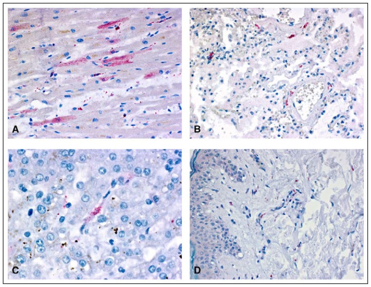
FIGURE.
Yellow fever virus antigens (red) detected after immunohistochemical staining in tissue samples from various organs* of a patient who died from yellow fever vaccine–associated viscerotropic disease — Oregon, September 2014
* Sample A: myocytes in heart; sample B: fibroblasts in vascular wall in lung; sample C: kupffer cell in liver; sample D: fibroblasts and histiocytes in skin. (Immunoalkaline phosphatase with naphthol fast-red substrate and hematoxylin counterstain. Original magnifications: A = ×400; B = ×100; C = ×400; D = ×100.)

