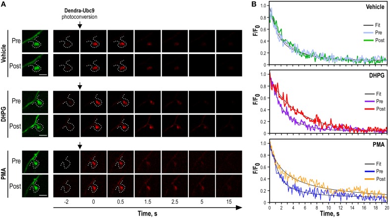
Figure 3.
Synaptic Dendra2-Ubc9 photoconversion experiments. (A) Time-lapse series of confocal images of photoconverted Dendra2-Ubc9 red fluorescence in spines before and after pharmacological stimulations. Images of spine before Dendra2-Ubc9 photoconversion are shown in green (left). Following the synaptic photoconversion, neurons were incubated with the mGlu5R agonist DHPG (50 μM), with the PKC activator PMA (2 μm) or in control (vehicle) solution for 10 min at 37°C as indicated. Dendra2-Ubc9 from the same spine was then photoconverted a second time and imaged for 20 s. Scale bar, 1 μm. (B). Representative sample paired recording traces of normalized fluorescence values obtained from individual photoconverted Dendra2-Ubc9 expressing spines before (Pre) and after (Post) vehicle, DHPG or PMA treatment as shown in (A). The thin black curves represent the corresponding fits. Note that some parts of this figure derived from Loriol et al. (2014).

