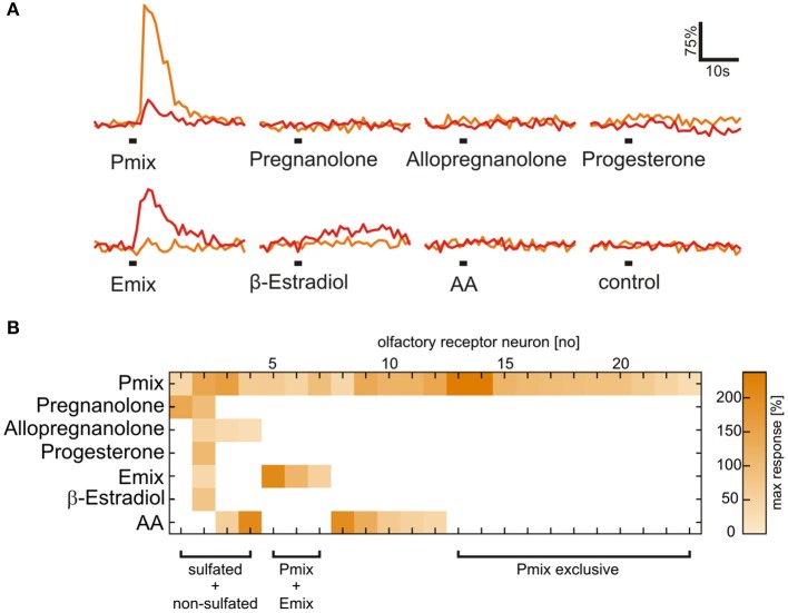
Figure 3.
Presence of a sulfate group is necessary for activation of most sulfated steroid-sensitive neurons. (A) Sulfated steroid-induced calcium responses of two neurons in the MOE (orange and red trace). Both neurons responded upon application of E and P sulfated steroids (200 μM), but were not sensitive to their non-sulfated analogs (200 μM). (B) Response matrix of MOE neurons sensitive to sulfated (E mix and P mix) and non-sulfated steroids (pregnanolone, allopregnanolone, progesterone, and β-estradiol; 23 cells, 2 slices). The majority of sulfated steroids-sensitive neurons (19 cells) did not respond upon application of non-sulfated steroids. Response intensity is coded by a color gradient. A mixture of amino acids (AA, 100 μM) was applied as a control for slice viability.

