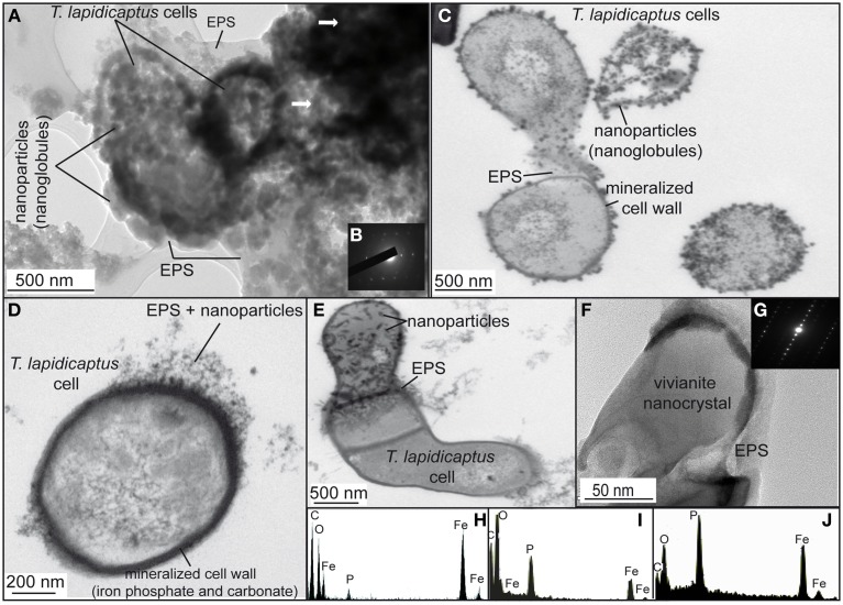
Figure 2.
TEM images of the bioprecipitates formed in T. Lapidicaptus anaerobic cultures. (A) The bioprecipitates are nanoparticles attached to T. lapidicaptus cell and its secreted EPS, respectively. (B) Electron diffraction pattern of the darker areas, more mineralized areas. (C) T. lapidicaptus cells with mineralized cell wall and covered by nanoparticles embeded in EPS. (D) Detail of T. lapidicaptus cell with mineralized cell wall. Note nanoparticles embedded in EPS. (E) Detail of three cells together with mineralized cell wall and EPS. The upper cell covered by nanoparticles embedded in EPS. (F) Elongated nanoparticle, vivianite nanocrystal, embedded in EPS. (G) Electron diffraction pattern of the nanocrystal (F). (H,I) EDX spectra of both dark and lighter mineralized areas (1A) composed of Fe-carbonate and phosphate, respectively. (J) EDX spectrum of nanoparticle (1E) composed of Fe-phosphate (vivianite).

