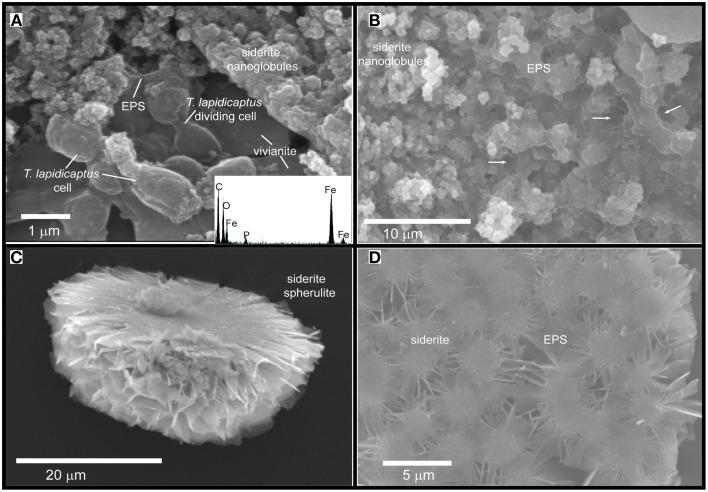
Figure 4.
SEM images of the Fe-carbonate precipitates from T. Lapidicaptus anaerobic cultures. (A) Siderite nanoglobules embedded in EPS and attached to mineralized dividing T. Lapidicaptus cells. Note the vivianite crystal attached to these cells. EDX spectrum of mineralized cell displaying C, O, Fe, and small peak of P. (B) Fe-carbonate nanoglobules (siderite) embedded in EPS and delimiting the bacterial cell contours (white arrows). These nanostructures display granulated texture. White arrows correspond to moulds of degraded bacteria (broken cells). (C) Broken microspherulite of siderite. (D) Detail of a siderite spherulite which formed by aggregation of nanoparticles.

