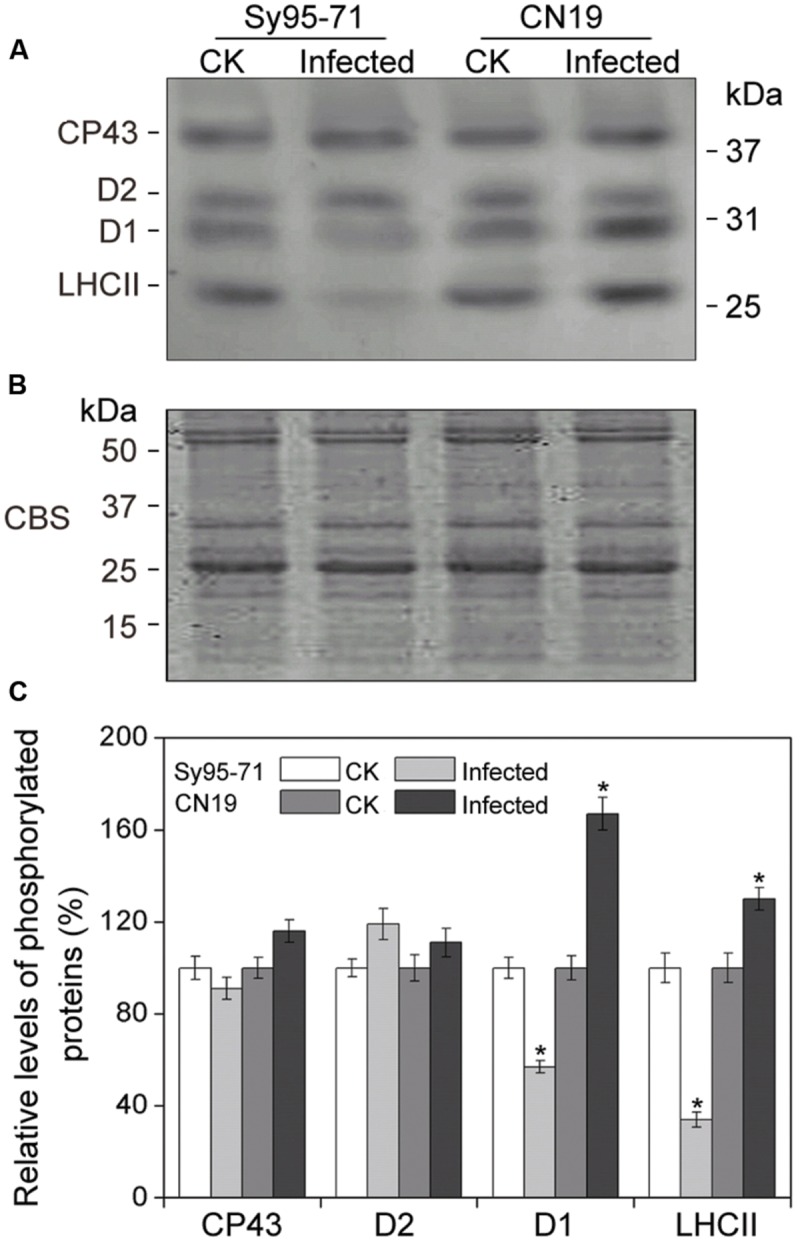
FIGURE 8.

Thylakoid protein phosphorylation after wheat stripe rust infection of the susceptible (Sy95-71) and resistant (CN19) wheat cultivars. Thylakoid proteins extracted from the inoculated and un-inoculated wheat plants were fractionated by SDS-PAGE in 12% acrylamide separation gel with 6 M urea. Immunoblot analysis of thylakoid membrane proteins was performed using anti-phosphothreonine antibodies (A). Loading was based on an equal amount of chlorophyll (1 μg chlorophyll). The SDS-PAGE results after Coomassie blue staining (CBS) are shown in the bottom panel (B). CK, un-inoculated wheat plants. (C) Quantification of immunoblot data. Results are presented relative to the amount of respective CK (100%). Asterisks indicates statistically significant differences at the P < 0.05 level. Values are means ± SD from three independent biological replicates.
