
Figure 4.
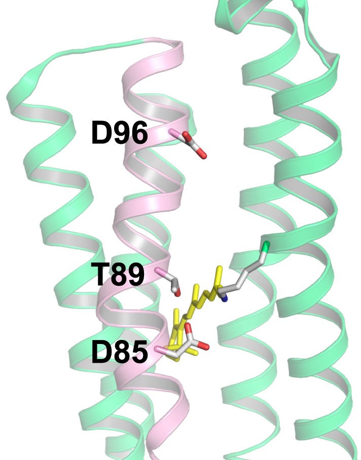
Structure of bacteriorhodopsin (BR) with the DTD motif (PDB: 1QM8, Takeda et al., 1998). The three amino acid residues of the motif (D85, T89, and D96) are located in the C-helix (pink), while other helices are shown in green. Among the seven helices, the A-helix and B-helix are removed to provide a clear view. In BR, D85, and D96 act as the H+ acceptor and donor to the Schiff base, respectively, and T89 forms a hydrogen bond with D85.