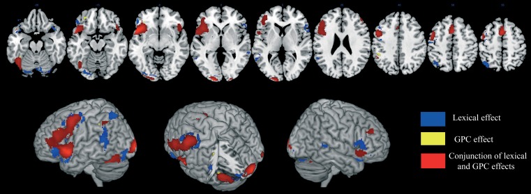
Figure 2.
Brain activation data. The cerebral areas that are specifically associated with lexical processing (in blue), sublexical processing (in yellow), and with both reading procedures (in red) are displayed on an anatomical template image (the “ch2better” template image in MRICron; Rorden and Brett, 2000).

