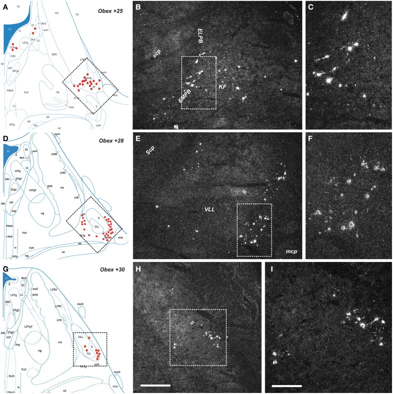
Figure 5.
Distribution of NPS mRNA-positive neurons in human pons. (A,D,G) Schematic drawings from the atlas by Paxinos et al. (2012), indicating distribution of NPS mRNA-positive cell bodies (red dots) at three different levels (Obex +25, +28, +30). All schematic drawings are presented in higher magnification in the Supplementary Material. Boxed area in (A,D,G) show (B,E,H), respectively. (B,C,E,F,H,I) Parabrachial cluster harbors numerous NPS mRNA-positive neurons. Note that the NPS neurons are distributed in the external medial/lateral parabrachial nuclei—Kölliker Fuse nucleus—lateral lemniscus region. The boxed area in (B), (E,H) show (C), (F,I), respectively. (A,D,G) are reproduced from Paxinos et al. (2012) with permission. Scale bars: 500 μm (H), applies to (B,E,H); 200 μm (I), applies to (C,F,I).

