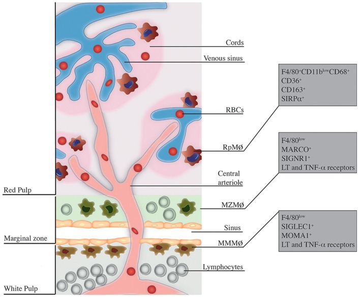
Figure 1.
Localization and phenotype of splenic MΦ subsets. This figure is a broad scheme of the positioning of RpMΦs, MZMΦs, and MMMΦs inside spleen and their respective phenotypic markers. RpMΦs (in red) are typically found within cords on the red pulp, allowing direct contact with RBCs and other blood cells/particles passing through venous sinuses. They are better defined by the concomitant expression of F4/80, CD11b (at low levels), and CD68 as well as other receptors that aid in their function. MZMΦs (in green) are found in the marginal zone (MZ) outer layer – they are also in direct contact with blood-borne particles. These cells express in their surface the molecules MARCO and SIGNR1 and other receptors that help in the uptake of blood-borne pathogens. Finally, the MMMΦs (in brown) reside within the inner layer of the MZ, in the contact with the white pulp. They are also specialized in blood-borne particle uptake and express surface markers such as SIGLEC-1 and MOMA-1.

