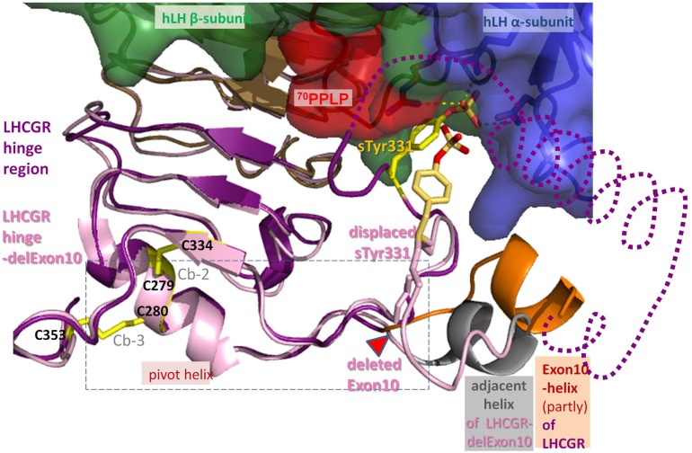
Figure 8.
Differences between hLH bound homology models of LHCGR hinge region and LHCGR hinge-delExon10. For LHCGR, the sulfated sTyr (yellow) of the hinge region (lilac) fits deep into the binding pocket of bound hLH between alpha- (surface blue) and beta-subunit (green, red adapted conformation for 70PPLP). For clarity, exon10-helix (orange) of LHCGR is cut open and the adjacent helix is omitted (dotted line). By contrast, the deletion in LHCGR-delExon10 (pale pink) causes displacement of the residues in the middle of the hinge region after the deletion position (red triangle). Subsequently sTyr331 is displaced and thus the interaction of the sulfation group with the hLH binding pocket is impaired. However, the deletion of 27 residues in LHCGR-delEx10 (contains exon10-helix) causes the polypeptide chain to contract, so that the adjacent helix (gray) is arranged in the same place as the exon10-helix (orange). Upon hormone binding, the signal is conveyed via the hinge region (lilac) through a hormone-induced movement of the pivot helix, the disulfide linked agonistic unit and the area prior to the exon10-helix, both of which are probably embedded (dashed box) in between the loops of the transmembrane domain of LHCGR.

