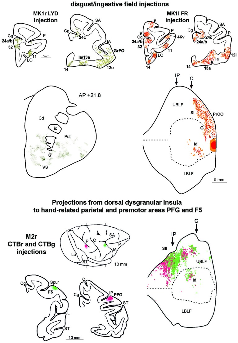
Figure 8.
Upper part left side: cortex and striatal representative coronal sections from MK1r LYD. In the drawings of the cortex coronal sections, dark green squares and light green circles correspond to the anterograde and the retrograde labeling, respectively. In the digitalized microphotograph of the striatum the extracted anterograde labeling is shown in green. Upper part right side: representative coronal sections and unfolded view of the LF from MK1l FR. In the draws of the sections and in the unfolded view of the LF red and orange circles corresponds to the anterograde and the retrograde labeling, respectively. Lower part: the location of the injection sites in hand-related premotor and parietal areas, F5 and PFG, are shown in draws of the 2D reconstructions of the hemispheres and in coronal sections. In the unfolded view of the LF the F5 and the PFG retrograde labeling is shown by green and red circles, respectively. Conventions and abbreviations as in Figures 2, 3, 4, 6.

