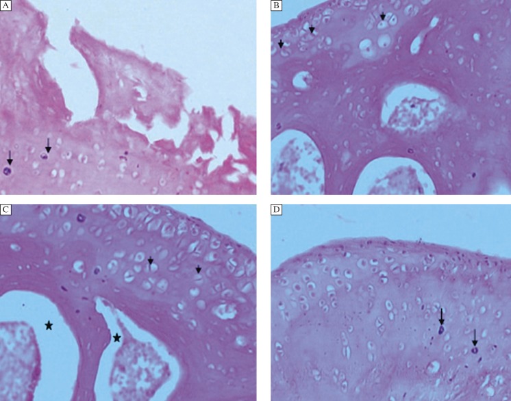
Fig. 4. Effect of treatment of diacerein after post 2 week IA injection.
A: Femorotibial joint 2 weeks post IA injection. The articular surface exhibits multifocal denuded cartilage with fissures extending deep into the deep layer. Large numbers of necrotic chondrocytes were seen (black arrow); (Grade 3.5, Stage 3).x200 B-Eight weeks after DC treatment was begun 2 weeks post IA injection. C, D: Twelve weeks after treatment was begun 2 weeks post IA injection. Regeneration of the cartilage is evident by the cell nest (small black arrow) within the articular cartilage. Note, in the subchondral region, the bone regeneration is poor. The hemopoitic tissue is being pushed to the center (*). Few necrotic chondrocytes are seen (down arrow)); (Grade 1.5-1, Stage 2-1). H & E,x400.

