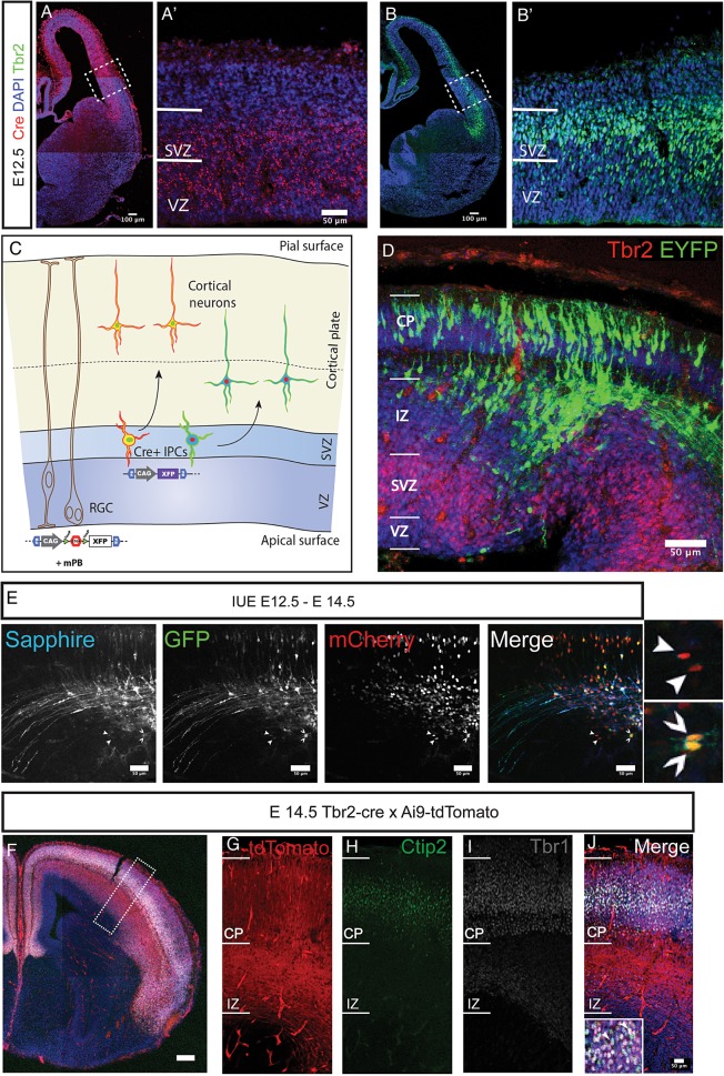
Figure 1.
Analysis of IPC derived neurons in the Tbr2Cre mouse with stable transfection of fluoroproteins and Ai9-tdTomato reporter mouse. (A,A′) Expression of the cre-recombinase in Tbr2Cre at E12.5 revealed with FISH. Cre mRNA is selectively expressed in the dorsal pallium and not in the subpallium in accordance with the known expression of Tbr2. Boxed region in A is shown with higher magnification in A′. (B,B′) Tbr2 immunoreactivity on an adjacent section to the one shown in A confirms the correlation between Tbr2 and Cre expression. Boxed region is shown with higher magnification in B′. (C) Schematic representation of the lineage analysis with stable transfection of CAG-STOP-floxed fluoroproteins using a constitutively expressing transposase plasmid by in utero electroporation. The constructs are taken up by the radial progenitors and passed on to IPCs where the recombination occurs leading to expression of fluorophores. (D) Single fluorophore (STOP-EYFP) transfection at E12.5 (collected at E14.5) revealed labeled cells in IZ, SP, and cortical plate, but not in VZ and SVZ, suggesting that the excision of the stop-floxed cassette occurs within the SVZ at the level of the Tbr2 positive IPCs (immunoreactivity for Tbr2 in red). (E) The method outlined in C can be extended to various CAG-STOP-floxed fluoroproteins to identify clonally derived cells. The shown example utilized 3 fluorophores (Sapphire, GFP, mCherry). Arrowheads indicate pairs of cells expressing the same combination of fluoroproteins. (F–J) Immunostaining sections from brains derived from breeding Tbr2Cre and Ai9-tdTomato mouse lines at E14.5 revealed that Ctip2 (H) and Tbr1 (I) immunoreactive neurons are derived through Tbr2 positive IPCs. Scale bars: A,B,F: 100 μm, rest are 50 μm.

