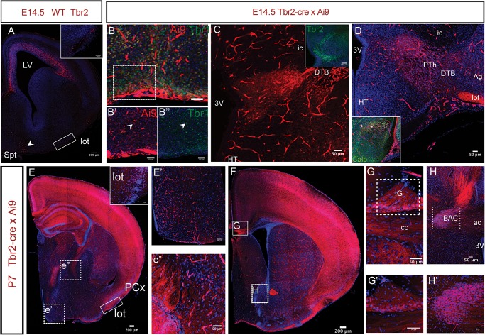
Figure 6.
Extracortical contributions of Tbr2+ IPCs. (A) Tbr2 immunoreactivity is detected at E14.5 in the septum (Spt, arrowhead) and in the lateral olfactory tract (lot). Inset shows expression in the lot from boxed area. LV, lateral ventricle. (B) Ai9+ (red) cells in the septal regions colocalise with Tbr1 (green) in the Tbr2Cre bred with Ai9-tdTomato. Tbr1 expression (B″, arrowhead) overlaps with Ai9 (B′, arrowhead) indicating glutamatergic nature of these cells. B′ and B″ are taken from the boxed region of an adjacent section. (C) Tbr2 is also expressed in the prethalamus (PTh) as indicated by Ai9+ cells at E14.5. Cells can be seen on either sides of the DTB. Inset shows expression of Tbr2 in the thalamic eminence. (D) On a more posterior section Ai9+ cells can be seen crossing the DTB and also in the lot at that level. Inset shows that some cells in the prethalamus coexpress calbindin (arrowheads). Abbreviations: 3V: 3rd Ventricle, HT: Hypothalamus, ic: internal capsule, Ag: amygdala, lot: lateral olfactory tract. (E) Coronal section from a brain derived from the breeding of Ai9-tdTomato and Tbr2Cre at P7 shows expression is maintained in the lot. Inset shows labeled cells in higher magnification in lot. PCx: piriform cortex. E′ and E″ show that cells in the hypothalamus and prethalamus also continue to express Ai9 at postnatal ages (taken from E at the sites of e′ and e″). (F) Cell of the indusium grisuem (IG; G,G′) and bed nucleus of anterior commissure (BAC; H,H′) express Ai9 in a P7 Tbr2Cre × Ai9 section. (G,H) show expression in a corresponding section. (G) Expression of Ai9 can be seen in the cells of the IG next to the midline and above the fibers of corpus callosum (also labeled by Ai9). Inset shows higher magnification. (H) Ai9 expression can be seen in BAC as well as in the anterior commissure. Inset shows higher magnification from the boxed region. Scale bars: a: 100 μm, EF: 200 μm while rest is 50 μm.

