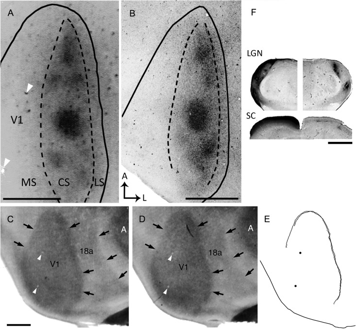
Figure 1.
Ipsilateral eye dominance domains in V1 demonstrated by transneuronal transport of WGA-HRP. (A,B) Tangential sections from 2 rats show dark patches of densely labeled axonal terminations. The black lines indicate the border of V1 as determined from the myelin pattern (see Materials and Methods). Dashed lines delineate the subdivision of V1 into medial (MS), central (CS), and lateral (LS) segments. (C,D) Myelin patterns from 2 tangential sections taken at the level of Layer 4 (∼500 and 540-μm deep, respectively) from case shown in A. Arrows indicate the borders of V1 and Area 18a as determined from the myelin pattern. An area of dense myelination corresponding to primary auditory cortex (A) is visible lateral to Area 18a. (E) Contours of Area V1 delineated from the myelin patterns in C and D using the filter “trace contour” in Adobe Photoshop (see Materials and Methods). These contours were used to draw the border of V1 in A. Black dots mark blood vessels that are also indicated in A,C,D (white arrowheads). Note that panel A is slightly rotated clockwise with respect to panels C–E. (F) Subcortical WGA-HRP labeling in case shown in A. Coronal sections through the dorsal lateral geniculate nucleus (LGN; top image), and superior colliculus (SC; bottom image) illustrating the contralateral (left), and ipsilateral (right) WGA-HRP labeling patterns. Dorsal is up. Scale bars = 1.0 mm.

