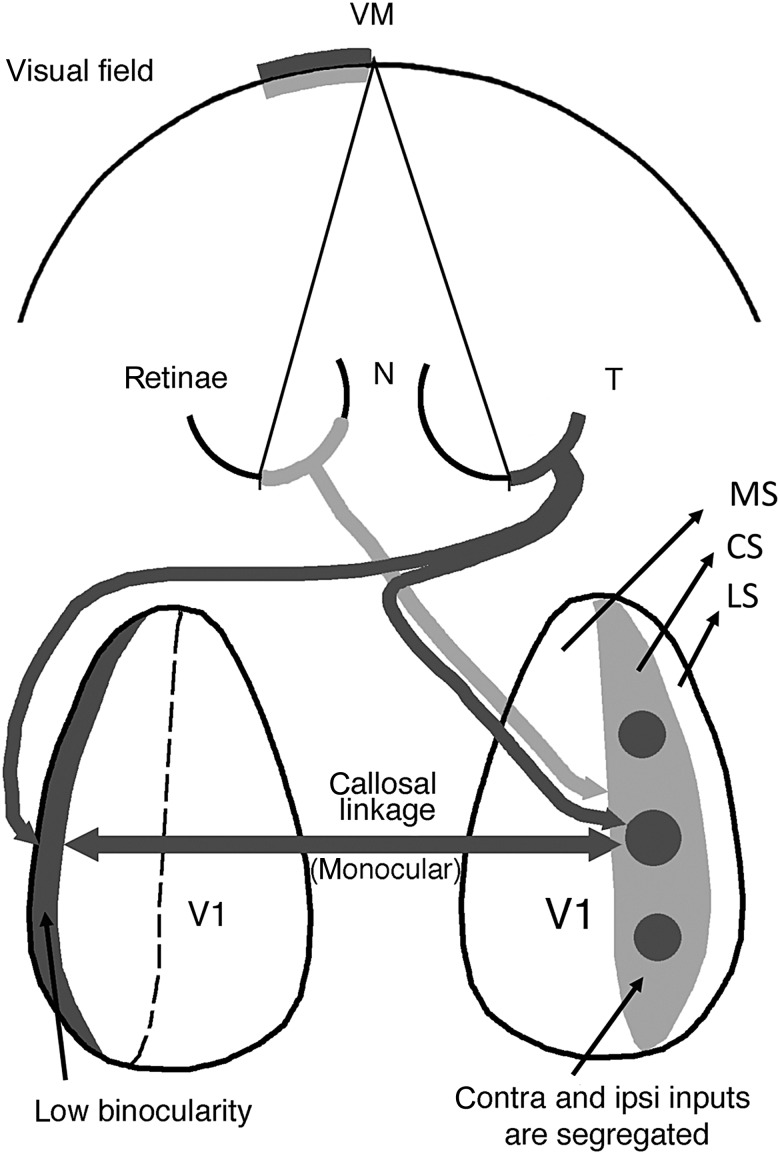
Figure 10.
Diagram relating the patterns of retinal projections to eye-specific domains in V1 and to the callosal linkages established between these domains. For simplicity only projections from the right temporal (dark gray) and left nasal (light gray) retinas are shown and the geniculate relay stations have been omitted. This diagram summarizes our finding that callosal connections correlate preferentially with ipsilateral eye domains in the CS (dark gray circular areas in the right CS) and with contralateral eye input in the LS (dark gray strip at left). The pathway colored light gray illustrates that input from the left nasal retina to the right CS is distributed preferentially to noncallosally connected regions (light gray regions) surrounding the ipsilateral eye domains, being therefore unable to convey ipsilateral eye input transcallosally to the left LS. This diagram helps explain our observation that LS in hooded rats is strongly dominated by the contralateral eye. Dashed line indicates the medial border of the CS. T, temporal; N, nasal; MS, medial segment; CS, central segment; LS, lateral segment. Diagram is based on our present results as well as previous studies of the point-to-point callosal connectivity in rat visual cortex (Lewis and Olavarria 1995; Olavarria and Hiroi 2003).

