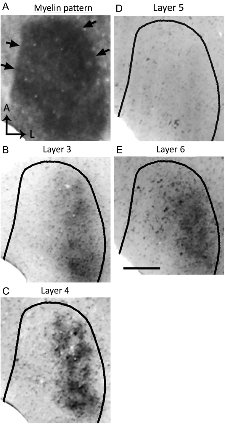
Figure 2.
Patterns of transneuronal WGA-HRP labeling at different depths in V1 following an injection of the enzyme into the ipsilateral eye. (A) Myelin pattern in section cut at ∼540 μm deep. Arrows indicate border of V1. (B–E) The pattern of WGA-HRP labeling was most distinct in sections through Layer 4 (C, 480 μm) and less distinct in sections through lower Layer 3 (B, 360 μm). The pattern was weak or not visible at the level of layer 5 (D, 720 μm), but it was again visible in sections through Layer 6 (E, 900 μm). The labeling pattern in sections through Layer 6 resembled the pattern observed in sections through Layer 4 suggesting that they are in register. Scale bar = 1.0 mm.

