Figure 6.
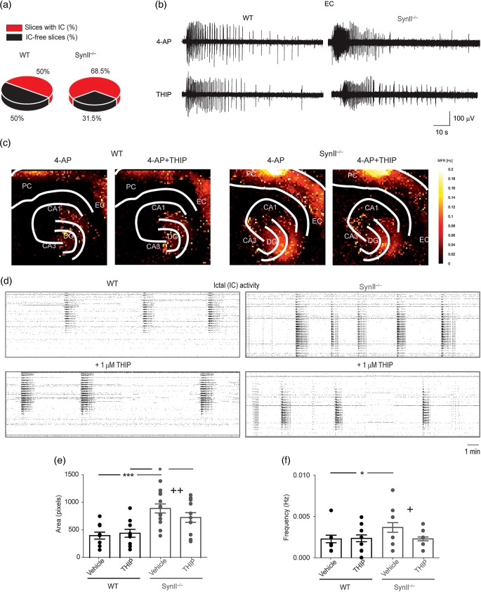
THIP rescues the epileptic-like IC activity in acute brain slices from Syn II−/− mice. (a) Occurrence of 4-AP-induced IC events in WT and Syn II−/− slices (P = 0.014, χ2 test with Yates correction). (b and c) Representative traces of IC activity induced by 100 μM 4-AP in the EC of WT and Syn II−/− slices before and after the administration of 1 μM THIP (b) and its regional distribution in the hippocampus and rhinal cortices (c) calculated from APS-MEA recordings (color-coded mean firing rate recorded from each 40-μm pixel electrode; see Materials and Methods). (d) Raster plots depicting highly synchronized IC activities recorded over the entire 4096 electrode field in WT (black frame) and Syn II−/− (gray frame) slices before and after THIP treatment. Note the reduction in the frequency induced by THIP in the Syn II−/− slices. (e and f) Bath application of 1 μM THIP reduces the epileptic area (e) and the increased frequency of IC events (f) in Syn II−/− slices to WT levels. *P < 0.05; ***P < 0.001 across genotype, +P < 0.05, ++P < 0.01, effect of treatment within genotype; two-way ANOVA followed by Bonferroni's post hoc test. DG, dentate gyrus; CA3, cornu ammonis 3; CA1, cornu ammonis 1; EC, entorhinal cortex; PC, perirhinal cortex.

