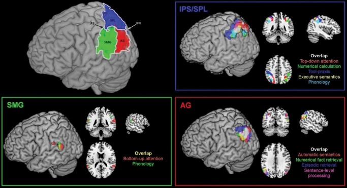
Figure 1.
Neuroanatomical location of the parietal cortex and its 3 major divisions; the results from the primary ALE analysis showing differential functional recruitment of IPS/SPL, SMG, and AG. Meta-analysis results were thresholded at FDR correction of P < 0.05 and a minimum cluster size of 100 mm³. For clarity, the images are masked to show data from the lateral parietal cortex only (see Supplementary Fig. 3 for whole-brain results).

