
Figure 5.
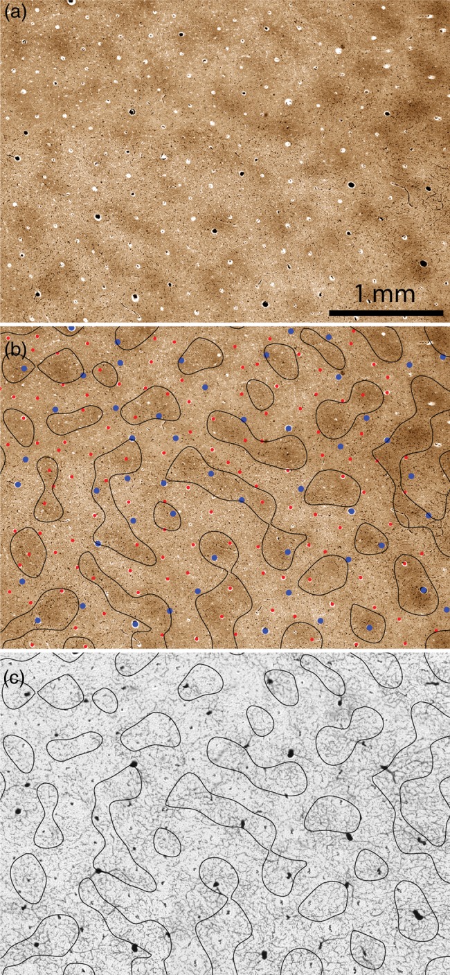
Relationship between microvascular lobules and CO patches. (a) CO patches from the boxed region in Fig. 2b. (b) Borders of the CO patches in (a) defined by thresholding the darkest one-third of pixels, with the blood vessels identified as either arterioles (red) or venules (blue) from the adjacent section in Fig. 3d. (c) Borders of the CO patches in (b) superimposed on the microvascular lobules shown in Fig. 3a. There is no coincidence between the vascular system and the CO patches.