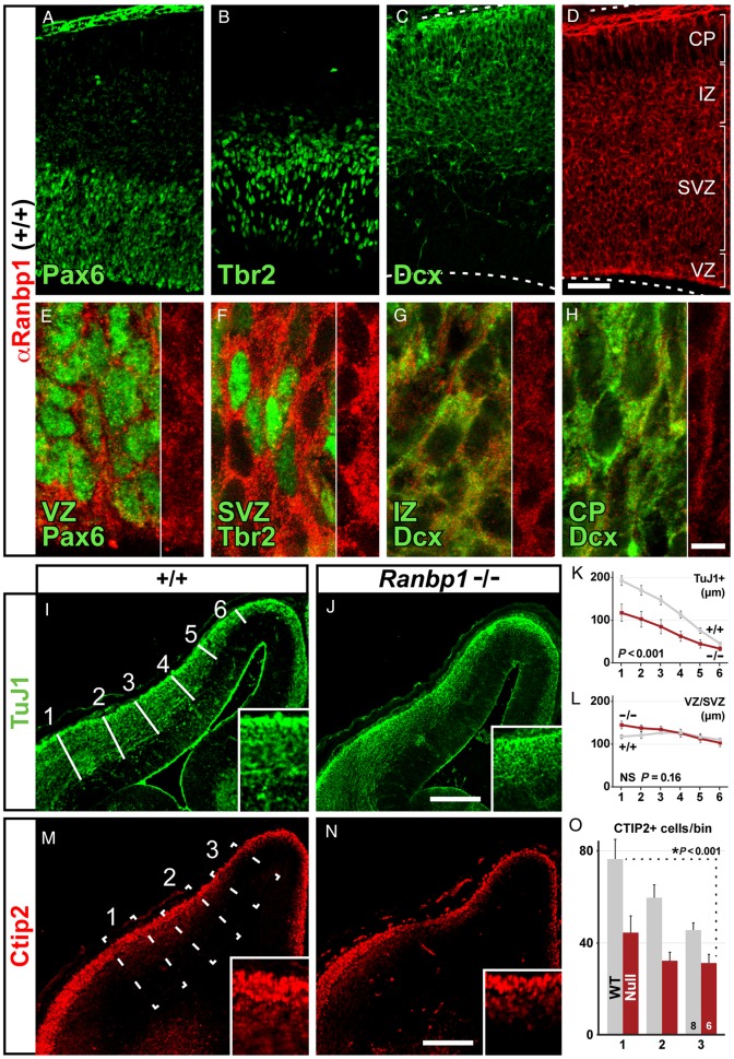
Figure 5.
Neurogenesis is disrupted in the Ranbp1−/− cortex. (A–H) Relative expression of Ranbp1 was assessed in E14.5 cortex by double labeling with markers for distinct progenitor subpopulations, including Pax6-expressing apical progenitors (A), Tbr2-expressing basal progenitors (C), as well as Doublecortin (Dcx)-positive intermediate zone (IZ) and cortical plate (CP) neuronal precursor cells, and immature neurons (C). Ranbp1 is expressed in all cortical layers (D), although high-magnification confocal micrographs indicate that levels are more robust in VZ (E) and SVZ (F), relative to IZ and CP. (I–L) Thickness of the TuJ1-expressing neuronal population is reduced in the Ranbp1 mutant, as measured at 6 evenly distributed locations across the cortical mantle (white lines, I). Quantification of the thickness of the TuJ1 layer shows reduction in Ranbp1−/− cortex (K; P < 0.001 by two-way ANOVA), whereas the thickness of the underlying VZ/SVZ layer is unchanged (L,P = 0.18). (M–O) Quantification of early generated neurons in 3 evenly distributed 150-µm-wide counting boxes (dashed lines, M) shows a reduction in the number of Ctip2-expressing neurons in the Ranbp1−/− cortex (O; P < 0.001 by two-way ANOVA). Scale bar for A–D = 50 µm; 5 µm for E-H, and 200 µm for I,J and M,N. Inset for I,J is ×2 magnification; ×3 magnification for M,N.

