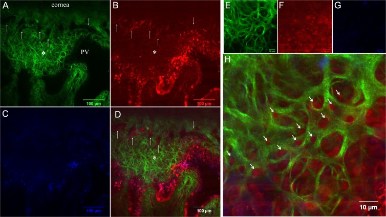
Figure 4.
Comparison of SHG (A, E) and AF (B, F) images at anterior limbal cribriform layer. (C, G) 4',6-diamidino-2-phenylindole (DAPI) and (D, H) merged images. (A–D) Images taken at 30 μm depth below the epithelium. The anterior cribriform layer is prominent in the SHG image (A, asterisk). Dark signal-void areas (arrows) in the SHG image (A) were occupied by AF-positive structure (B). DAPI was used to counterstain nucleus (C). Merged image (D) clearly demonstrates the orientation of collagen and vessels. (E–H) Magnified images of anterior limbal cribriform layer. Arrows indicate the AF positive structure occupying each honeycomb space of anterior limbal cribriform layer. Colors are pseudocolors. Images were taken from sample no. 4.

