
Figure 6.
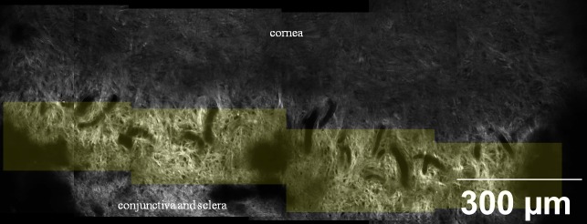
A montage of SHG images taken at 45 μm depth below the epithelium. Yellow tint represents anterior limbal cribriform layer. This structure is clearly distinguished from adjacent structures, such as cornea and sclera. Cribriform area spanned 360 degrees around the limbus. Images were taken from sample no. 1.