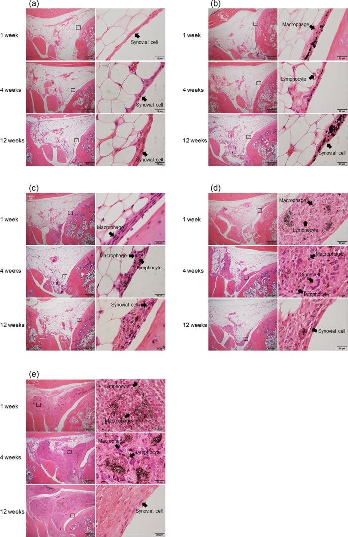
Figure 1. Analysis of the effects of a single intra-articular dose of MWCNTs using hematoxylin-eosin staining.

(a) Histological images of the control group captured at 1, 4, and 12 weeks after physiological saline injection. Superficial tissue and deep adipose cells of synovial tissue were normal at all time points, and no inflammatory response was observed. (b) Histological images of 0.3 mg carbon black (CB) injection. After 1 week, CB invaded the synovial surface and was incorporated by macrophages, and a mild inflammatory response was observed primarily in the lymphocytes. CB was not observed in adipose tissue. After 4 weeks, the inflammatory response was improved, although CB was still incorporated in macrophages. At 12 weeks, the inflammatory response was resolved. (c) Histological images of 0.003 mg MWCNTs into the knee joint. After 1 week, MWCNTs invaded the synovial surface, which became mildly thickened. MWCNTs were incorporated into macrophages, and a mild inflammatory response was observed primarily in lymphocytes. After 4 weeks, the inflammatory response was alleviated, although MWCNTs were still incorporated in macrophages. At 12 weeks, the surface was covered with normal synovial tissue. (d) Histological images of 0.03 mg MWCNTs into the knee joint. After 1 week, MWCNTs had invaded the deep synovial tissue, and inflammatory cells (macrophages and lymphocytes) had replaced a portion of the adipose tissue. MWCNTs were aggregated and incorporated into macrophages. After 4 weeks, the inflammatory area was reduced, and a multinucleated foreign body giant cell made of fused macrophages was observed. After 12 weeks, the inflammatory response was resolved, and granulation tissue was formed in the normal synovial cells. (e) Histological images of 0.3 mg MWCNTs. MWCNTs invaded a larger area than that of the 0.03 mg group; however, no severe inflammatory response was observed. The inflammatory response was reduced after 4 weeks and resolved after 12 weeks. Granulation tissue was formed in a wider area than that in the 0.03 mg group; however, the synovial surface was covered with normal cells.
