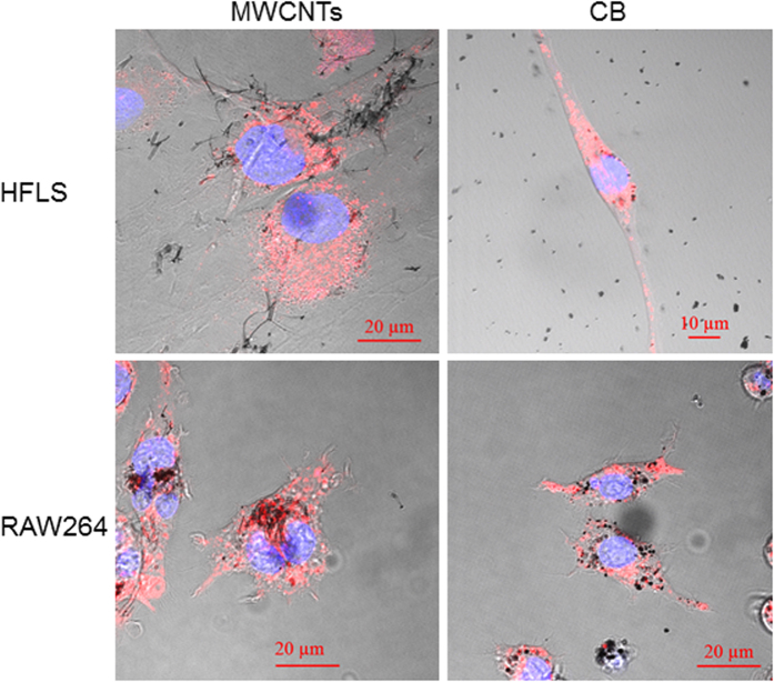
Figure 3. Incorporation of MWCNTs into cells.
Human fibroblast-like synoviocytes (HFLSs) and RAW264 cells were cultured and exposed to either 10 μg/mL MWCNTs or CB for 24 h. Both HFLSs and RAW264 cells exhibited incorporation of MWCNTs and CB, with RAW264 cells showing increased incorporation per cell. Both MWCNTs and CB were observed in lysosomes of HFLSs and RAW264 cells. Blue: nucleus, Red: lysosome.

