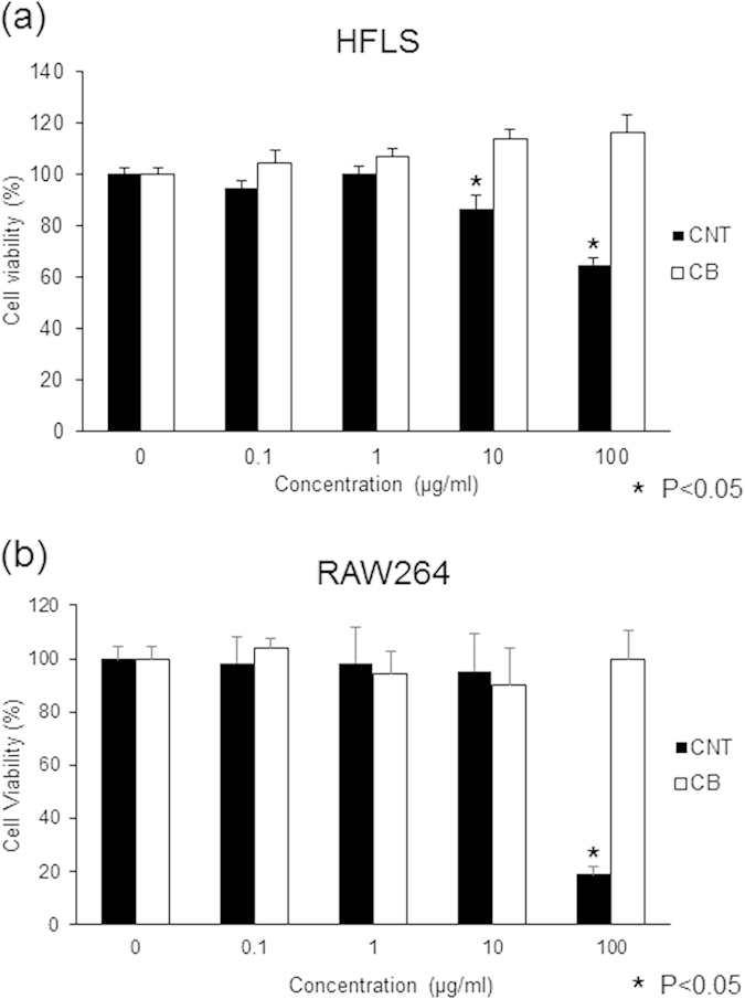
Figure 4. Proliferation of HFLSs and RAW264 cells exposed to MWCNTs.

Cultured HFLSs and RAW264 cells were exposed to 0, 0.1, 1, 10, or 100 μg/mL of MWCNTs or CB. After 24 h of exposure, Alamar Blue staining was performed, and cell numbers were counted 4 h later. (a) In HFLS cells, CB did not result in inhibition of proliferation at any concentration. MWCNTs resulted in a dose-dependent decrease in cell proliferation at 10 and 100 μg/mL at rates of 86% and 64% that of the control, respectively. The difference was statistically significant. (b) For RAW264 cells, CB did not result in inhibition of proliferation at any concentration. When treated with 100 μg/mL MWCNTs, cell proliferation was inhibited at a rate of 18% that of the control. The difference was statistically significant.
