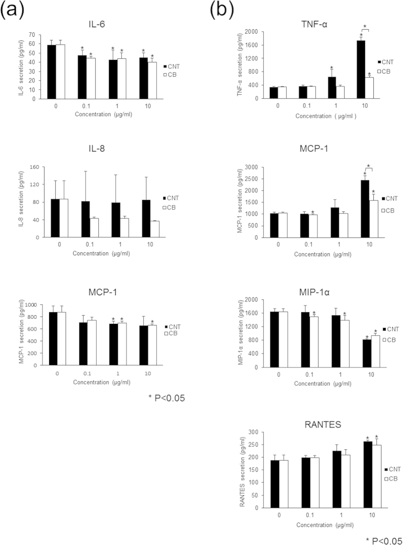
Figure 5. Cytokine and chemokine secretion from HFLSs and RAW264 cells exposed to MWCNTs.
Cultured HFLSs and RAW264 cells were exposed to 0, 0.1, 1, or 10 μg/mL MWCNTs or CB. After 24 h of exposure, detection and measurement of cytokines and chemokines were performed. (a) HFLSs secreted IL-6, IL-8, and MCP-1. IL-6 secretion was reduced by both MWCNTs and CB exposure, but the dose-dependent effects were not clear. IL-8 was unchanged by MWCNTs and showed a decreasing tendency with CB, without dose dependency, and the difference was not significant. MCP-1 secretion was significantly reduced by 1 μg/mL MWCNTs and 1 or 10 μg/mL CB, but the effects were not dose dependent. (b) RAW264 cells secreted TNF-α, MCP-1, MIP-1α, and RANTES. TNF-α secretion was significantly increased by 1 or 10 μg/mL MWCNTs and 10 μg/mL CB, and MWCNTs resulted in a larger increase than did CB. MCP-1 secretion was increased by 10 μg/mL MWCNTs and CB, and MWCNTs resulted in a larger increase than did CB. MIP-1α secretion was significantly decreased by 10 μg/mL MWCNTs and was significantly decreased in response to CB in a dose-dependent manner. RANTES secretion was significantly increased by both 10 μg/mL MWCNTs and CB.

