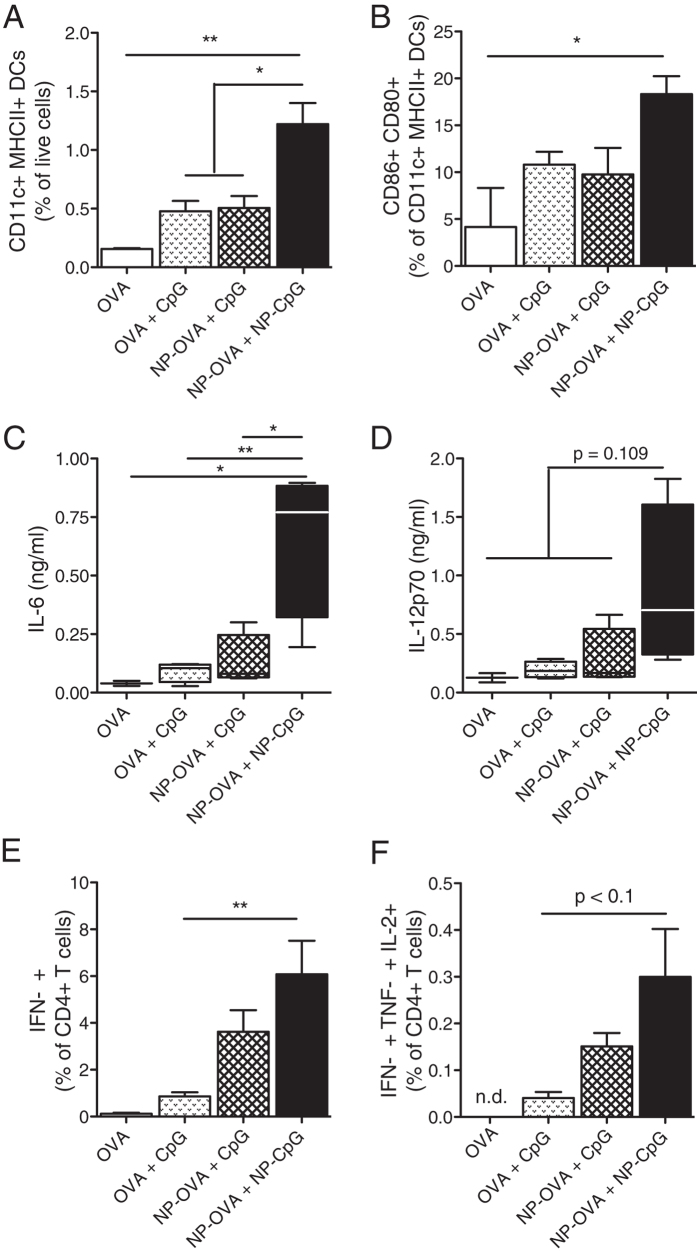
Figure 1. Pulmonary NP-CpG activates lung-resident DCs and leads to Th1 immunity in lungs.
Mice received 5 μg OVA and 2 μg CpG (free or NP-conjugated) via the pulmonary route and (A–D) sacrificed 24 h later or (E,F) immunized again on day 14 and sacrificed on day 19. (A) Frequencies of all dendritic cells (DCs, CD11c+ MHCII+) in the lungs, as percentages of live cells. (B) Co-expression of the activation markers CD86 and CD80 (as percentages of CD11c+ MHCII+ DCs). (C) IL-6 and (D) IL-12p70 concentrations in the bronchoalveolar lavage (BAL) as determined by ELISA. (E) IFN-γ+ T cells and (F) polyfunctional (IFN-γ+ TNF-α+ IL-2+) T cells in lungs after restimulation with OVA (as percentages of CD4+ T cells). Data show mean ± SEM from two independent experiments, 7 mice per group (2 mice in OVA group). *P < 0.05, **P < 0.01.

