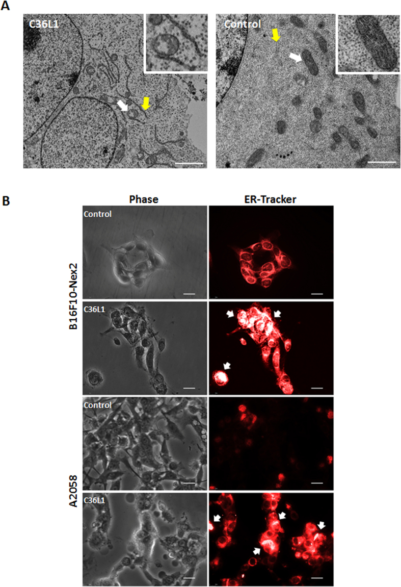
Figure 2. Effects on mitochondria and endoplasmic reticulum (ER).
(A) Transmission electron microscopy (TEM) of B16F10-Nex2 previously incubated with C36L1 at 1.2 nmoles/103 cells for 18 h. White arrows, normal and altered vacuolated mitochondria; yellow arrow, normal and condensed ER; Scale bar represents 2 μm; (B) C36L1 induces ER condensation of cells of B16F10-Nex2 and A2058 cells. Tumor cells were previously incubated with peptide C36L1 at 5 nmoles/103 for 3 h and further stained with fluorescent ER-tracker. The morphology of ER was documented by fluorescence microscopy (Nikon Instruments, Inc, Melville, NY). White arrow indicates high condensation area. Scale bar represents 20 μm.

