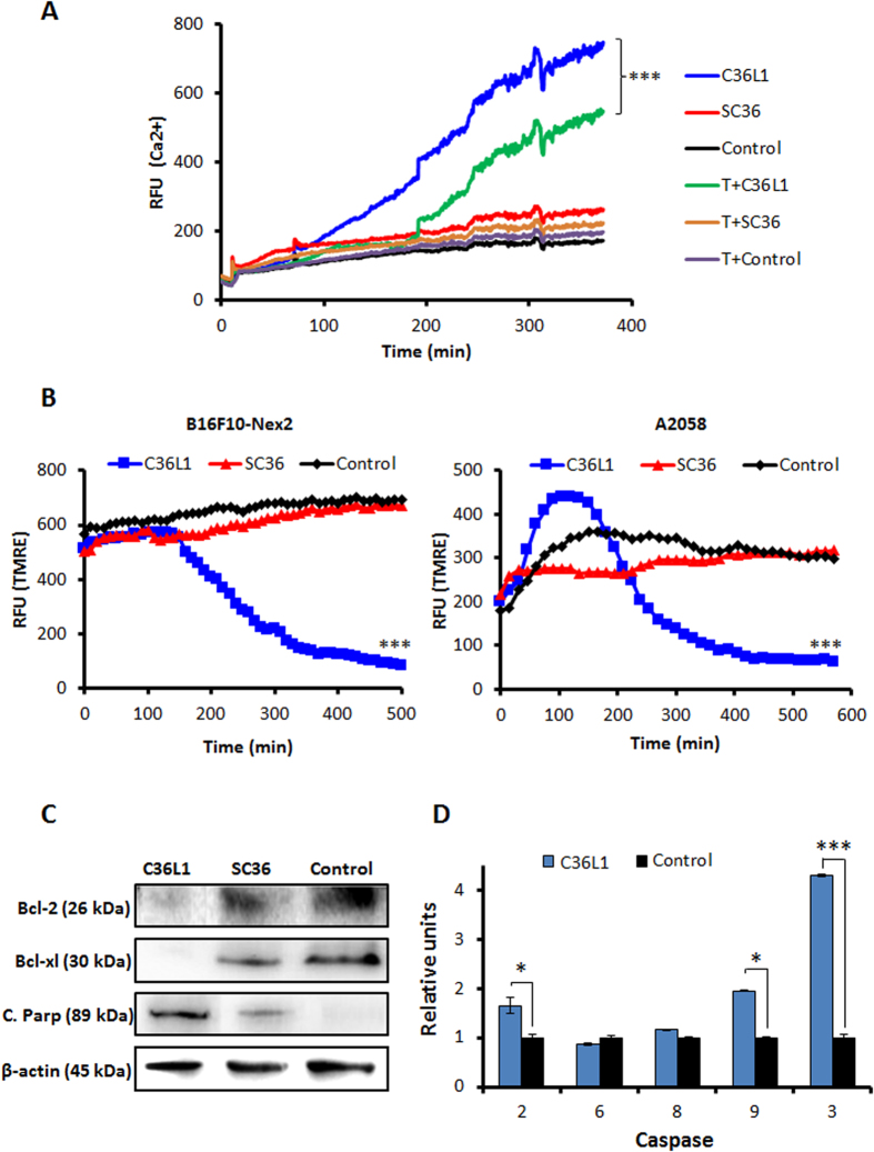
Figure 3. Functinoal effects of C36L1 in ER and mitochondria.
(A) C36L1 induces ER Ca2+ release in B16F10-Nex2 cells, added at 5 nmoles/103 cells for 6 h (***p < 0.001, in relation to T (thapsigargin) + C36L1 group); (B) C36L1 induces collapse of the mitochondrial transmembrane potential (∆ψm) in B16F10-Nex2 and A2058 cells during treatment at 5 nmoles/103 cells for 6h (***p < 0.001, in relation to untreated control); (C) Lysates from B16F10-Nex2 cells, previously treated with peptides at 5 nmoles/103 cells for 18 h at 37 °C were analyzed for prosurvival and proapoptotic proteins by Western blotting with the following antibodies: Bcl-2, Bcl-xl and C. Parp (Cleaved Parp). Anti β-actin was used as total protein loading control (***p < 0.001, in relation to untreated control). (D) Caspase-2, −6, −8, −9 and −3 activity in B16F10-Nex2 cells incubated with C36L1 at 1.2 nmole/103cells for 2 h (*p < 0.05; **p < 0.01; **p < 0.001, in relation to untreated control).

