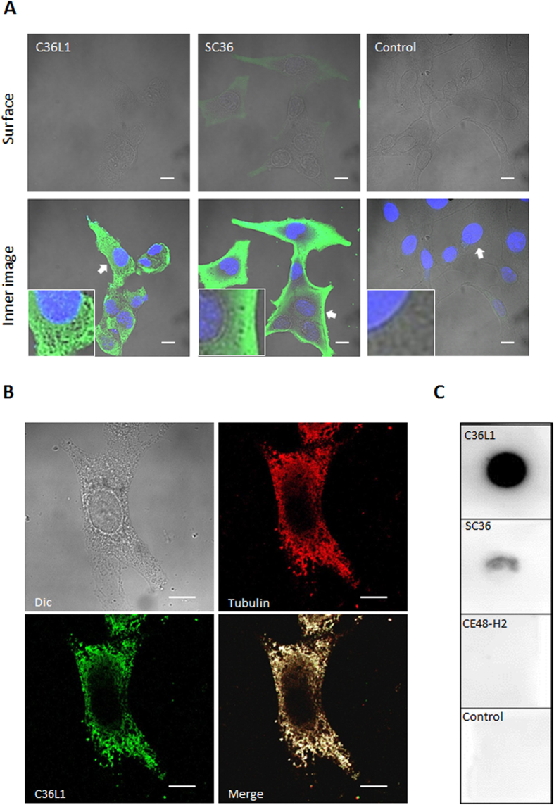
Figure 5. Peptide internalization and reactivity with microtubules.
(A) C36L1 and SC36 internalization in B16F10-Nex2 cells incubated with 1.5 nmoles/103 cells of biotinylated peptides for 30 min. Images were acquired by confocal fluorescence microscopy (CFM). Scale bar represents 10 μm; (B) C36L1 colocalizes with microtubules of B16F10-Nex2 cells analyzed by CFM (magnification, ×100). Scale bar represents 10 μm; (C) C36L1 binds to tubulin present in the lysate of B16F10-Nex2 cells. Dot-blots were performed by coating the nitrocellulose membranes with 3 μg of each peptide C36L1, SC36 and CFE48-H2 (inactive CDR peptide control) in 3 μL vehicle (Milli-Q water). Experimental and control dot-blots were performed as described in methods in the following order: (a) C36L1/tubulin; (b) SC36/tubulin; (c) CE48-H2/tubulin; (d) Control (vehicle). Detection was made with anti-α-tubulin mAb.

