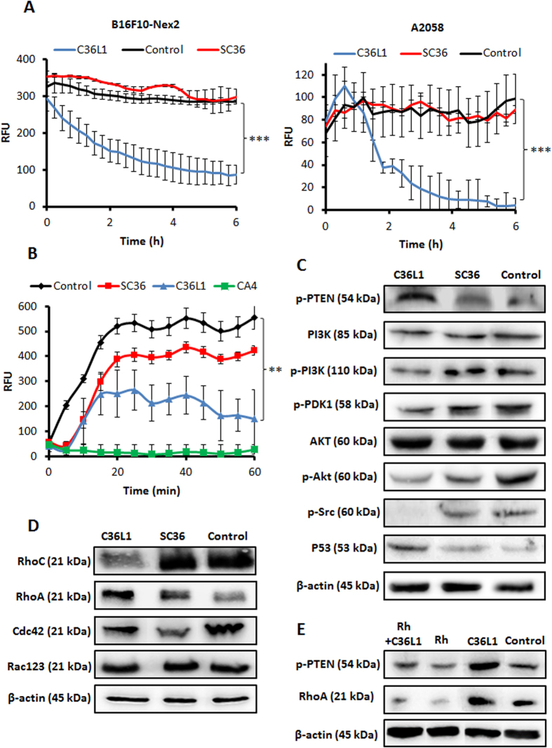
Figure 6. Microtubule de-stabilization and cell signaling.
(A) B16F10-Nex2 and A2058 cells expressing baculovirus-transduced fluorescent microtubules were incubated with C36L1 and SC36 at 5 nmoles/103 cells and microtubule integrity was quantified in real time as described in Methods; (B) C36L1 and SC36 (15 nmoles/well) were incubated with tubulin and polymerization kinetics was measured. CA4 (7.5 nmoles/well) were used as positive control (**p < 0.01; **p < 0.001, in relation to untreated control); (C) Lysates from B16F10-Nex2 cells, previously treated with peptides at 0.25 nmole/103 cells for 18 h at 37 °C, were analyzed by Western blotting. The following antibodies to p-PTEN (S380), total PI3K, p-PI3K (p110α), p-PDK1 (S241), total Akt, p-Akt (S473), p-Src (T416) and total p53, were used; (D) The Rho-GTPase family proteins were also evaluated with antibodies to RhoC, RhoA, Cdc42 and total Rac123; (E) Lysates from B16F10-Nex2 cells, previously treated with Rh 50 μM for 12 h and then incubated with C36L1 at 0.4 nmol/103 cells for 18 h at 37 °C, were analyzed by Western blotting for p-PTEN (S380) and RhoA levels. β-actin was used as total protein loading control.

