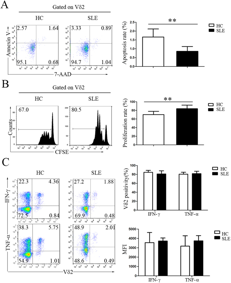
Figure 2. The decrease in peripheral Vδ2 T cells in SLE patients was not caused by either increased apoptosis or decreased proliferation.
(A) Fresh PBMCs from healthy controls (HC) and SLE patients were stained with an anti-Vδ2 mAb, 7-AAD and Annexin V. The data were gated for Vδ2 T cells. The frequency of 7-AAD and Annexin V double-positive labeling represents apoptosis. (B) Fresh PBMCs were labeled with CFSE and expanded using an immobilized anti-pan-TCRγδ mAb. (C) Intracellular staining for IFN-γ and TNF-α in Vδ2 T cells. **p < 0.01.

