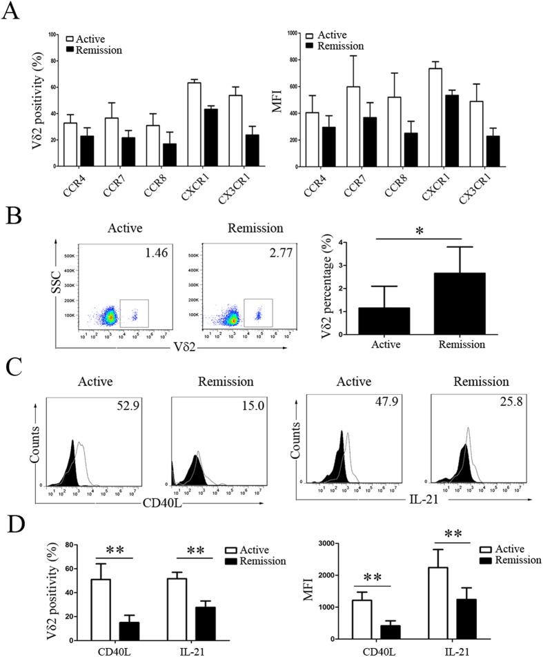
Figure 7. Repopulation of peripheral Vδ2 T cells with downregulated levels of chemokine receptors and costimulatory factors in SLE patients in remission.
Comparison of (A) the positivity and MFI of individual chemokine receptor expression on Vδ2 T cells, (B) the percentage of peripheral Vδ2 T cells, and (C,D) the positivity and MFI of CD40L and IL-21 expression in Vδ2 T cells between new-onset active and remitted SLE patients. The filled graphs represent isotype controls, and the unfilled graphs represent CD40L or IL-21staining. *p < 0.05, **p < 0.01.

