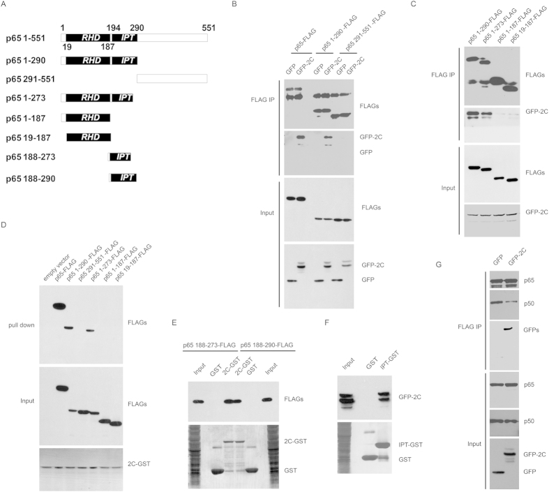
Figure 2. IPT domain of p65 associated with 2C.
(A) The diagram of p65 truncations. Numbers indicated the amino acid position. (B) EV71 2C interacts with p65 1-290aa. 293T cells transfected with 2C and truncation constructs of p65 were analyzed by coimmunoprecipitation and Western blots using indicated antibodies. (C) EV71 2C interacts with 1–273 and 1–290 of p65. 293T cells transfected with 2C and truncation constructs of p65 were analyzed by coimmunoprecipitation and Western blots using indicated antibodies. (D) 1–273 and 1–290 of p65 interacts with 2C. 2C-GST immobilized on glutathione-Sepharose beads were incubated with lysates from 293T cells transfected with p65-FLAG or truncated p65-FLAG plasmids. The bound proteins were subjected to Western blots using indicated antibodies. (E) 188–273 and 188–290 of p65 interacts with 2C. 2C-GST or GST immobilized on glutathione-Sepharose beads were incubated with lysates from 293T cells transfected with indicated truncated p65-FLAG plasmids. The bound proteins were subjected to Western blots using indicated antibodies. (F) p65 IPT interacts with 2C. IPT-GST or GST immobilized on glutathione-Sepharose beads were incubated with lysates from 293T cells transfected with GFP-2C plasmid. The bound proteins were subjected to Western blots using indicated antibodies. (G) 2C inhibits p65/p50 dimerization. 293T cells transfected with p65, p50, 2C or GFP constructs were harvested and analyzed by coimmunoprecipitation and Western blots using indicated antibodies.

