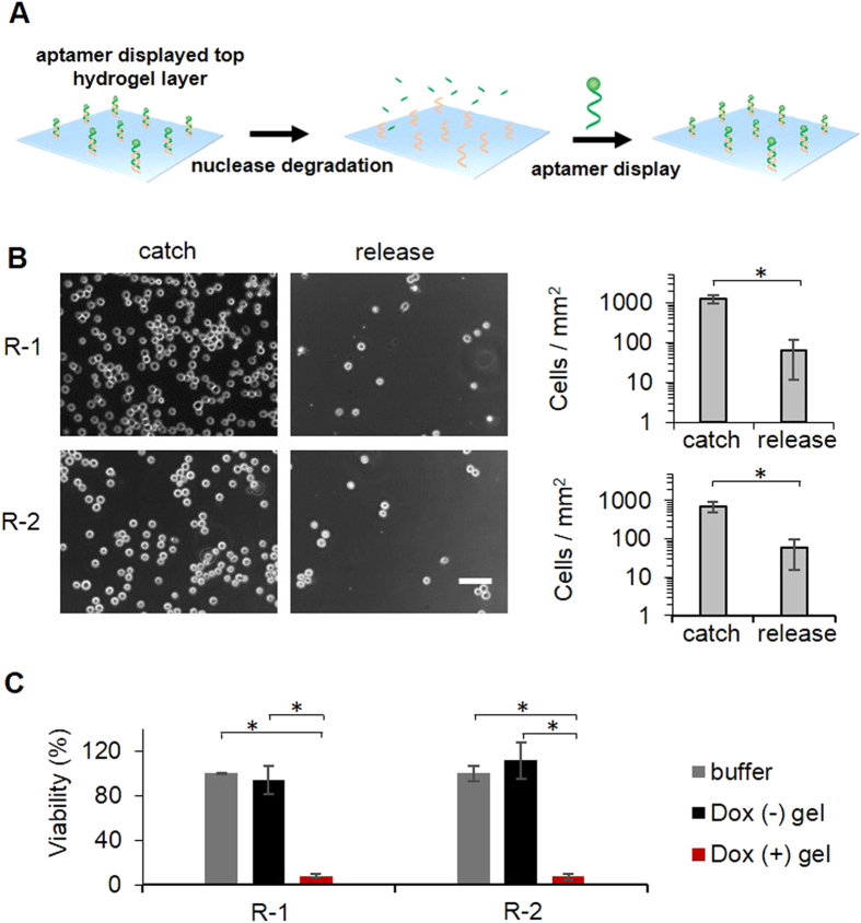
Figure 8. Evaluation of sustained cell catch and killing.
(A) Schematic illustration of alternate aptamer display and nuclease degradation on the top layer hydrogel. ID1 is nondegradable owing to the full modification of its backbone whereas unmodified CD1 is degradable. (B) Microscopic images and quantitative analysis of CCRF-CEM cell catch and release in a two-round test. Scale bar: 50 μm. (C) Analysis of cell viability. *P < 0.05; n = 3.

