
Figure 3. Effects of C-Pc on H-astrocytes for 24 h.
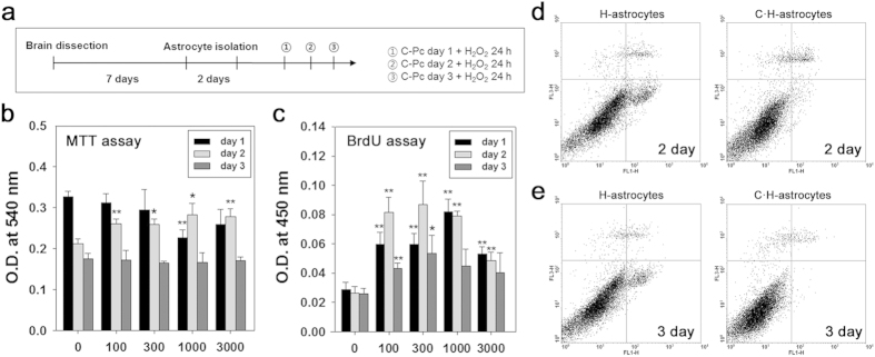
(a) Astrocytes isolation and treatment of C-Pc and H2O2 were implemented according to the schedule. (b) Viability and (c) proliferation of astrocytes were assessed by MTT and BrdU assay, respectively. The C-Pc concentrations were 0, 100, 300, 1000, and 3000 ng/ml and the treatment times were 1, 2, and 3 day. The apoptosis rate (%) of H-astrocytes and C·H-astrocytes (300 ng/ml of C-Pc) was determined by Annexin-V and PI staining and flow cytometry at (d) day 2 and (e) day 3. Data are presented as the means ± SEMs. The asterisk denotes significant difference against day 1 sample at each concentration. *p < 0.05 and **p < 0.01