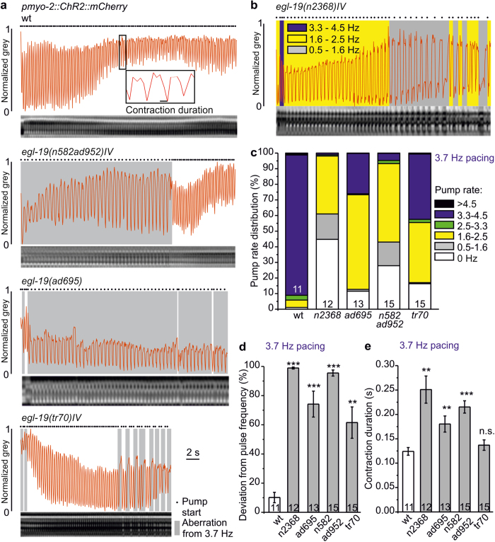
Figure 7. egl-19 mutants exhibit highly arrhythmic pumping despite steady optical pacing, as shown by kymographic analyses.
(a) Kymographic analyses of egl-19 mutant animals compared to wild type (wt), as indicated, during 3.7 Hz pacing (35 ms light pulses, over 30 s). Kymographs are shown below in each panel; the derived normalized grey values are shown as red traces. Periods of observed pumping deviating from 3.7 Hz are shaded grey. Pump starts are shown by black dots. (b) Pump rate distribution at 3.7 Hz pacing in a egl-19(n2368)IV animal represented by shaded areas (blue: 3.3–4.5 Hz, yellow: 1.6–2.5 Hz, grey: 0.5–1.6 Hz. (c) Group data, distributions of pump rates (black: >4.5 Hz, blue: 3.3–4.5 Hz, green: 2.5–3.3 Hz, yellow: 1.6–2.5 Hz, grey: 0.5–1.6 Hz, white: 0 Hz) in the indicated egl-19 mutants, compared to wild type (wt), obtained from animals paced at 3.7 Hz (n = 11–15 animals). (d) Mean deviation (±s.e.m.) of observed pharynx pumping from the pacing frequency, for the indicated fraction of stimulation period, given in % for wild type and the indicated egl-19 alleles (n = 11–15 animals). (e) Contraction duration (mean ± s.e.m.) of the animals analyzed in d. Statistically significant differences in (d,e): t-test with Bonferroni correction (***P < 0.001; **P < 0.01; *P < 0.05).

