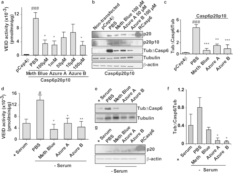
Figure 4. Methylene blue, azure A, and azure B inhibit active Casp6 in HCT116 cells and human primary neurons.
(a) Casp6 VEIDase enzymatic activity in pCep4β vector- and pCep4βCasp6p20p10-transfected HCT116 cells treated with PBS, methylene blue (Meth blue), azure A, and azure B for 2 hours. Data represent the mean and SEM of 4 independent experiments. Statistical differences were evaluated with one-way ANOVA (p ≤ 0.0001) and post hoc Bonferroni test comparing PBS or phenothiazine-treated cells with pCep4β (#) or pCep4βCasp6p20p10-transfected HCT116 cells (*). ###p < 0.001, *p < 0.05. (b) Western blot analysis of transfected- or non-transfected HCT116 cells shown in panel a for active Casp6p20 subunit (p20), full length Casp6 lacking its pro-domain; Casp6p20p10 (p20p10), Tubulin-cleaved by Casp6 (TubΔCasp6), full length Tubulin and β-actin. (c) Densitometric quantification of the levels of Tub∆Casp6 in three independent experiments as shown in panel b. Data represent the mean and SEM of 3 independent experiments. Statistical evaluation conducted as described in a. ###p < 0.001, **p < 0.01, ***p < 0.001. (d) Casp6 VEIDase enzymatic activity in serum-deprived primary human neuron cultures treated with 100 μM methylene blue, 50 μM azure A, and 10 μM azure B or PBS for 2 hours. Data represent the mean and SEM of 4 independent experiments. Statistical differences were conducted with a one-way ANOVA (p < 0.041) followed by a post hoc Bonferroni test comparing PBS or phenothiazine-treated cells with serum-treated (+serum; #) or serum-deprived PBS-treated cells (PBS; *) #p < 0.05, *p < 0.05, **p < 0.01). (e) Western blot analysis of neuronal protein extracts shown in panel d for levels of Tub∆Casp6 and full length tubulin. (f) Quantification of the levels of Tub∆Casp6/total tubulin shown in panel e. Data represent the mean and SEM of 3 independent experiments. Statistical evaluations by one-way ANOVA (p = 0.0315) followed by post hoc Dunnett’s test comparing to serum-treated (no significant difference) and serum deprived PBS-treated neurons (*p < 0.05). (g) Western blot analysis of the p20 active subunit of Casp6 (p20) and β-actin in proteins from serum-deprived human neurons.

