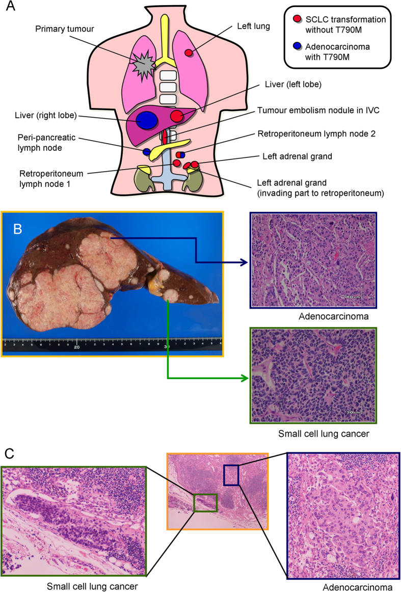
Figure 1. Anatomical and pathological examination of gefitinib-refractory metastatic lesions of the patient.
(A) Schema of the metastatic lesions available. There were no viable tumour cells in the primary lung tumour. Red lesions indicate adenocarcinoma histology, and all adenocarcinoma lesions harboured the T790M mutation. Blue lesions indicate SCLC histology, and none of the SCLC lesions had the T790M mutation. One retroperitoneum lymph node possessed both the adenocarcinoma component with a T790M mutation and the SCLC component, independently. (B) Macroscopically, there were two types of tumours in the liver. Lesions in the right lobe consisted of adenocarcinoma histology. Lesions in the left lobe showed SCLC histology. (C) Detail of the retroperitoneum lymph node that possessed both the adenocarcinoma and SCLC components is shown.

