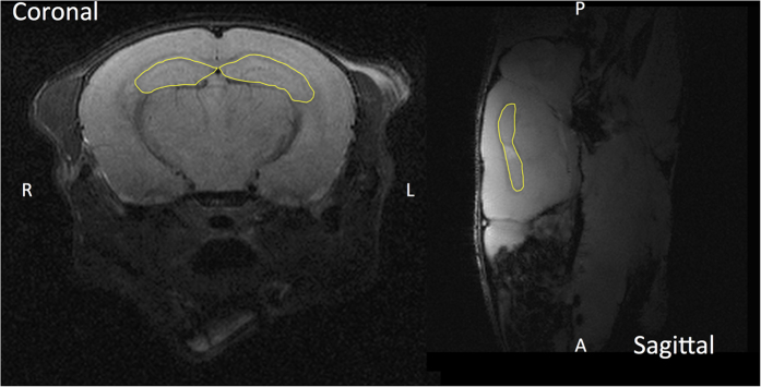
Figure 1. Anatomical outline of the mouse hippocampi.

Mouse hippocampi highlighted as a yellow contour on coronal and sagittal T1-weighted magnetic resonance images, obtained using a 9.4 T small animal magnetic resonance imager. The right/left and anterior/posterior directions are indicated in respective panels.
