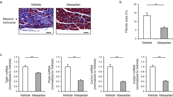
Figure 2. Fibrosis in cryoinjured skeletal muscle of irbesartan-treated mice.
(a) Histological sections with Masson’s trichrome staining of TA muscles in mice treated with irbesartan or vehicle at 14 d after cryoinjury. Scale bars, 50 μm. (b) The percent area of fibrosis in Masson’s trichrome staining of TA muscles in mice treated with irbesartan or vehicle at 14 d after cryoinjury (n = 5, in each group). Data are presented as mean ± SEM. **P < 0.01. (c) The mRNA levels of Tgfb1, Postn, Col1a1, and Col3a1 in TA muscles of mice treated with irbesartan or vehicle at 10 d after cryoinjury (n = 8, in each group). Data are shown as fold induction over vehicle (mean ± SEM). **P < 0.01.

