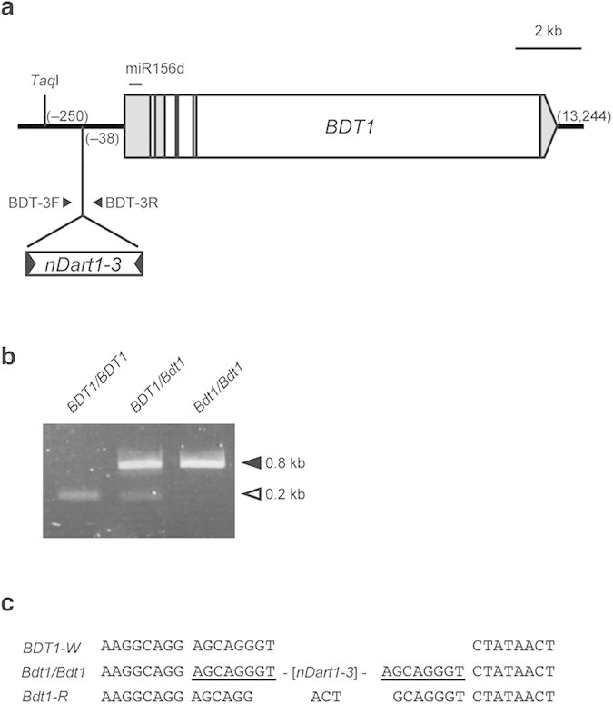
Figure 3. Characterization of the Bdt1 allele.

(a) Structure of the BDT1 gene carrying the nDart1–3 insertion. Shaded and white boxes represent BDT1 exons and introns, respectively. The horizontal line above the box represents the position where mature miR156d is transcribed. The numbers in parentheses represent the nucleotide positions from the transcription initiation sites of full-length cDNA AK073452. The small horizontal arrowheads indicate positions of the primers used for PCR amplification to characterize the insertions of nDart1–3 (Fig. 2b). (b) Genotyping of WT and Bdt1 plants by PCR analysis. The filled and open arrowheads indicate PCR-amplified bands with and without nDart1 insertion, respectively. (c) Sequence of the footprints generated by nDart1–3 excisions. The WT, homozygous Bdt1, and germinal revertant alleles are indicated by BDT1-W, Bdt1/Bdt1, and Bdt1-R, respectively. Target site duplication generated by nDart1–3 insertion at the Bdt1 locus is underlined.
