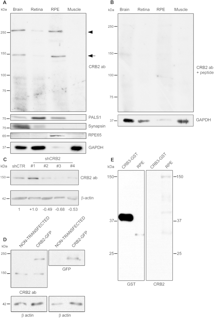
Figure 2. Specificity analyses for the CRB2 antibody and CRB2 expression in the RPE.
(A) The CRB2 antibody detects two bands with approximate molecular weights of 150 kDa (arrow) and 260 kDa (arrowhead) in brain, retina and RPE, but not in muscle. A member of the Crumbs complex, PALS1, is also expressed in the same tissues. The synapsin protein, only present in brain and retina lysates, and the expression of RPE65, only found in RPE, rule out any cross-contamination between retinal and RPE lysates. (B) In the peptide competition assay, the two bands detected at 150 and 260 kDa with the CRB2 antibody disappear. GAPDH was used as loading control. (C) CRB2 antibody detects a band of approximately 150 kDa in N2A cells. The intensity of this band decreases in cells transfected with three different shRNAs of a set of four (shCRB2) when compared with shRNA control (shCTR) transfected cells. The numbers indicate the CRB2 increase/decrease values relative to the shCTR. (D) CRB2-GFP fused protein overexpressed in HEK293 cells (CRB2-GFP) is detected as a band of 180 kDa by CRB2 and GFP antibodies but not in non-transfected HEK293cells. The 150 kDa band is also detected by the CRB2 antibody in both transfected and non-transfected HEK293 cells. β-actin was used as loading control. (E) CRB3 protein fused to GST is detected wit GST antibody at 40 kDa but not with CRB2 antibody. RPE tissue was used as a control of CRB2 antibody viability.

