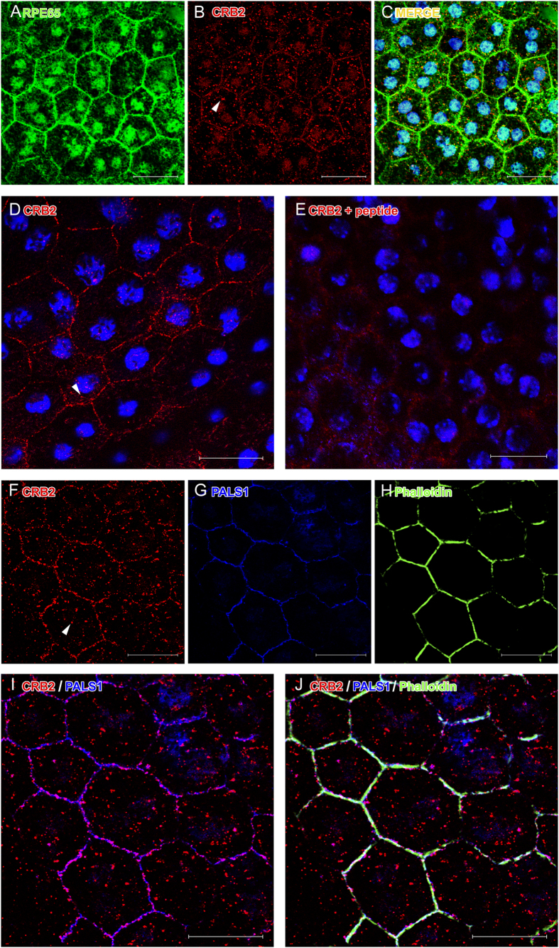
Figure 3. CRB2 detection by IF on RPE flatmounts.
RPE65 (A) is expressed in RPE cells together with CRB2 (B). (C) Merged images of RPE65 and CRB2 labeling and nuclei in blue. (D) CRB2 is located in the cell membrane of RPE cells. (E) The CRB2 labeling disappears after the peptide competition assay. Nuclei are stained in blue in (D,E). Triple IF showing CRB2 (F), PALS1 (G) and phalloidin (H) in the cell membrane. CRB2 and PALS1 colocalize in the cell membrane (I) together with phalloidin (J). Arrowheads in (B,D,F): scattered punctate labeling for CRB2 in the cytoplasm. Scale bars: 20 μm.

