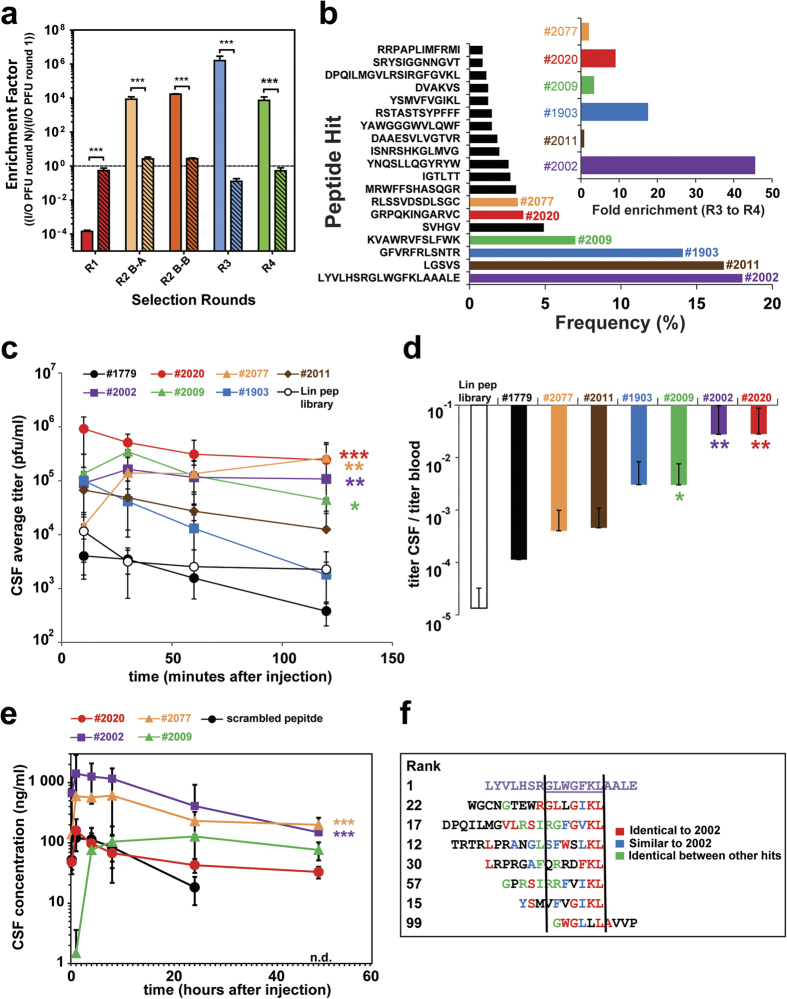
Figure 3. Validation of CSF enriched phage-displayed peptides and CSF transport of biotinylated lead peptides conjugated to streptavidin payload.
(a) Calculated enrichment factors based on injected (input = I) phage titers (PFU) and determined CSF phage titers (output = O) over all four rounds (R1-R4). The enrichment factors for the last three rounds (R2-R4) were calculated by the comparison with the previous round and the first round (R1) with the wt data. Open bars are CSF and stipple bars are plasma. (***p < 0.001, based on students t-test). (b) List of the most enriched phage peptides, ranked based on their relative proportion to all CSF collected phages after the 4th selection round. The six most frequent phage clones are highlighted in colors, assigned with a number and their enrichment factors between the 3rd and 4th selection round (insert). (c,d) The six most enriched phage clones of the 4th round, the empty phage and the stock phage peptide library individually analyzed in the CSF sampling model. CSF and blood samples were collected at the indicated time points. (c) Equal amounts of 6 candidate phage clones (2 × 10¹⁰ phages/animal), empty phages (#1779) (2 × 10¹⁰ phages/animal) and the stock phage peptide library (2 × 10¹² phages/animal) were tail vein i.v. injected in at least 3 CM cannulated animals each. The CSF pharmacokinetics of each injected phage clone and phage peptide library is displayed over time. (d) Display the average CSF/blood ratios of all recovered phage/ml over the sampling time. (e) Four synthesized peptide leads and a scrambled control attached through their N-terminal biotin to streptavidin (tetrameric display) and subsequently injected (tail vein i.v., 10 mg streptavidin/kg) in at least three cannulated rats (N = 3). CSF samples were collected at the indicated time points and the streptavidin concentration was measured by an anti-streptavidin ELISA in the CSF (n.d. = not detected). (*p < 0.05, **p < 0.01, ***p < 0.001, based on ANOVA test). (f) Comparison of the amino acid sequence of the most enriched phage peptide clone #2002 (purple) with other selected phage peptide clones from the 4th selection round. Identical and similar stretches of amino acids are color coded.

