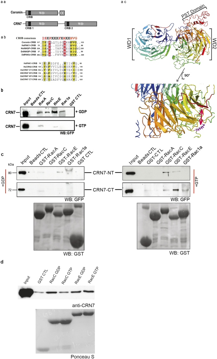
Figure 1. The CRIB domains of CRN7 and Rac GTPase interactions of CRN7.
(aa) Domain structure of coronin and CRN7. (ab) Sequence alignment of the CRIB domain in coronin and CRN7 and selected proteins. The consensus sequence is shown above the alignment. The position within the proteins is indicated. (ac) Model of CRN7. The 3D structure was predicted as described11. The CRIB domains are in shown red. A magnification of the region containing the CRIB domain and the putative F-actin binding site in WD2 is shown below. The conserved K residue is indicated in a stick model in magenta. (b) CRN7 interacts primarily with GDP-bound Rac. GST and GST-Rac proteins loaded with GDP or GTPγS were bound to Glutathione Sepharose beads and incubated with lysates of corB− cells expressing GFP-CRN7 WT. The bound proteins were analyzed by western blotting. Probing was with GFP specific mAb K3-184-2. (c) The N- and the C-terminal half of CRN7 interact with Rac GTPases. The precipitated proteins were detected with mAb K3-184-2, the GST-fusions with polyclonal GST-specific antibodies. GST control (GST CTL) and beads control (Beads CTL) are shown. (d) CRN7 interacts directly with Rac proteins. Bacterially expressed GST-CRN7-NT encompassing the PST-domain was cleaved from the GST part and used in pull down assays with GST-Rac GTPases. The precipitated proteins were analyzed in western blots. CRN7-NT was detected with mAb K67-31-5.

