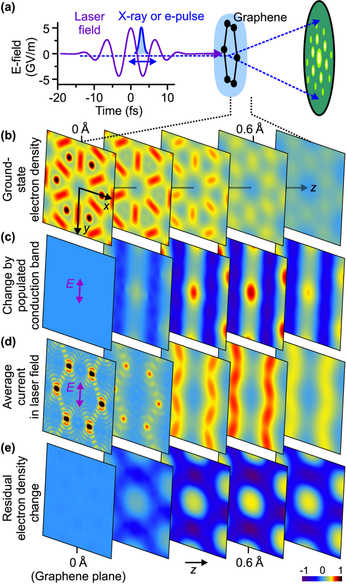
Figure 1. Real-space imaging of electronic motion in condensed matter.
(a) A few-femtosecond electron or X-ray pulse (blue) probes how the charge density in a sample, here graphene, changes under influence of an intense mid-infrared laser pulse (violet). The intensities of Bragg spots (green) are measured for a series of delays between the pulses. (b) Ground-state electron density of graphene’s valence-band electrons in several planes parallel to the sample; the distance to the centre of the atomic layer varies from z = 0 in steps of Δz = 0.2 Å. (c) Change of the electron density caused by excitation from the top 2 eV of the valence band below the Fermi energy to the bottom of the conduction band. (d) Time-averaged magnitude of the atomic-scale electric current. (e) Change of the microscopic electron density after the interaction with the laser pulse. In panels (b–e), the coordinates x and y span the range ±2.1 Å.

