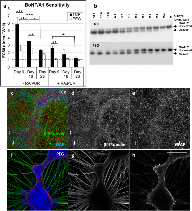
Figure 3. Morphologies and BoNT/A1 sensitivity for iPSC-NSCs cultured on TCP or PEG hydrogel surfaces.
A comparison of iPSC-NSCs cultured on PEG hydrogels (PEG) or PLO/LAM coated TCP surfaces (TCP), differentiated for 5 days with (+) or without (−) RA/PUR and matured for 9, 16, or 23 days. (a) BoNT/A1 sensitivities (EC50, Mean ± S.D., 3 replicate experiments) for iPSC-NSCs cultured on TCP or PEG surfaces. The EC50 value is defined as BoNT activity (Units/Well) required to reach half the maximum response for SNAP-25 cleavage, where 1 U is equivalent to the mLD50 determined using an in vivo mouse bioassay (dashed line). Statistical significance was determined using a one-way ANOVA (alpha = 0.05) followed by a Tukey test to compare individual means (Multiplicity adjusted P-values: *P ≤ 0.05; **P ≤ 0.01; ***P ≤ 0.001) See Supplementary Table S1 for all sample comparisons. (b) Western blot data showing SNAP-25 cleavage for iPSC-NSCs that were differentiated for 5 days (with RA/PUR) and matured for 23 days and then treated with serial dilutions of BoNT/A1 (Units/Well). (c–h) Immunofluorescence imaging illustrating (c,f) βIII-tubulin (neurons, green), GFAP (glial, red), and DAPI (nuclei, blue) expression and single channel grayscale images for (d,g) βIII-tubulin, and (e,h) GFAP. Human iPSC-NSCs were differentiated (+RA/PUR) and matured (23 days) on (c–e) PLO/LAM treated TCP and (f–h) PEG hydrogels with 50% non-degradable SH-PEG-SH crosslinks and 3 mM CRGDS. Scale Bars: 250 μm.

